")
")
Partner lab price: 15.66 €/h
Public and private lab price: 55.41 €/h
Public and private lab price: 55.41 €/h
Associated technologies:
- 2D imaging
- 3D imaging
Brief description:
Upright widefield epifluorescence microscope ideal for 2D or 3D acquisitions followed by deconvolution, for fixed samples between slide and coverslip. Apotome module for optical sectioning.Multi-dimensional acquisition
Acquisition software: ZEN Blue 3.2

Objectives:
5X EC Plan Neofluar 0.15 NA
10X EC Plan Neofluar 0.3 NA
20X Plan Apochromat 0.8 NA
40X EC Plan Neofluar 1.3 NA oil
63X Plan Apochromat 1.4 NA oil
100X alpha Plan Apochromat 1.46 NA oil
5X EC Plan Neofluar 0.15 NA
10X EC Plan Neofluar 0.3 NA
20X Plan Apochromat 0.8 NA
40X EC Plan Neofluar 1.3 NA oil
63X Plan Apochromat 1.4 NA oil
100X alpha Plan Apochromat 1.46 NA oil
Camera: Hamamastu Orca flash 4.0 - B&W, 16-bits, 2048x2048 pixels, pixel size @6.5 µm
- - -
Excitation source: HXP120V Lamp
- - -
Filter cubes:
Hoechst
GFP
Cy3
Texas Red
Cy5
FluorGold
Excitation source: HXP120V Lamp
- - -
Filter cubes:
Hoechst
GFP
Cy3
Texas Red
Cy5
FluorGold
- - -
Type of acquisition:
Automated on a slide: XYZ, multichannel, mosaic
Customizable protocol
Type of acquisition:
Automated on a slide: XYZ, multichannel, mosaic
Customizable protocol
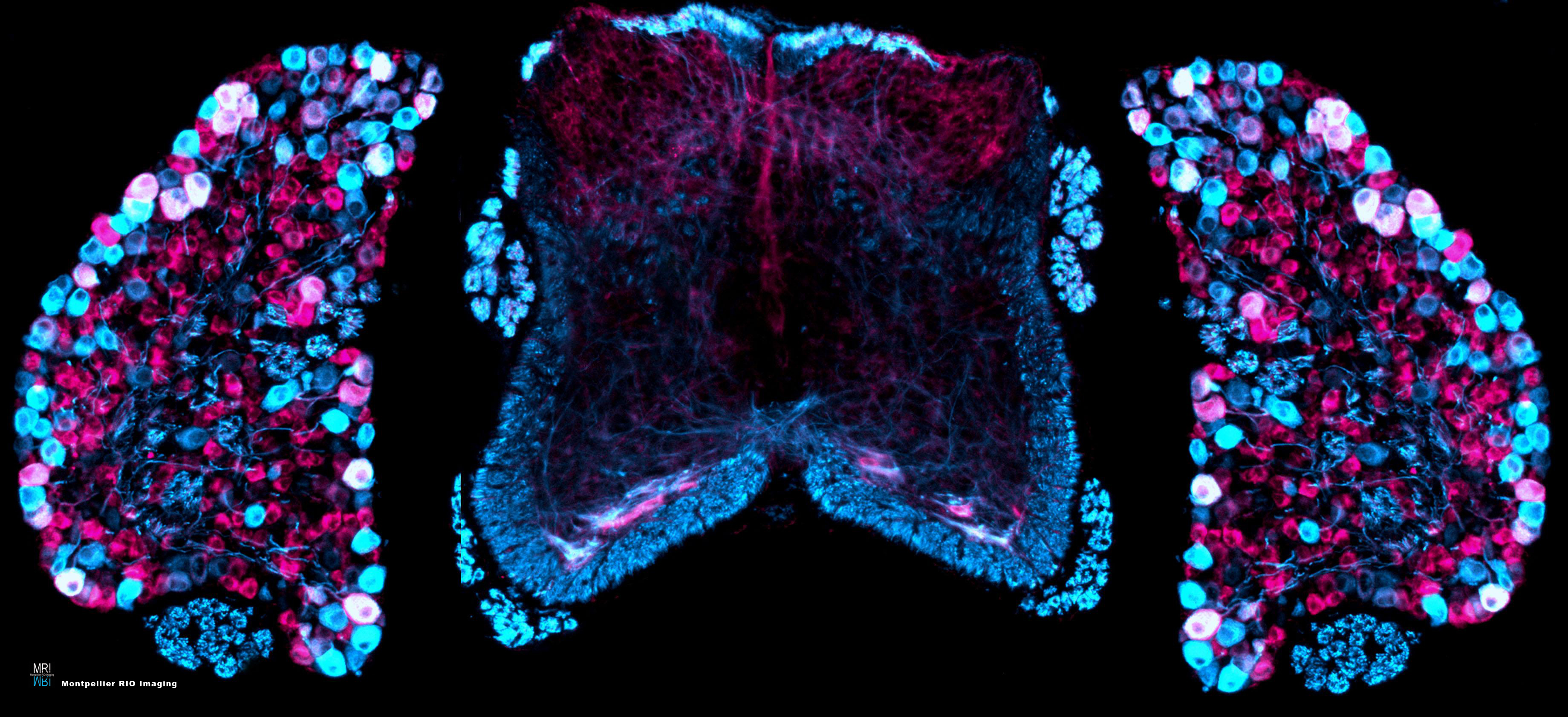
Mouse spine - MRI 2007 Image Contest 2nd prize
Transverse section of mouse spine at P0 stage, with spinal cord in the center and dorsal root ganglia on either side: Ret (Magenta), NF200 (Cyan).
Steve Bourane, Institute for Neurosciences of Montpellier (INM), Montpellier
Transverse section of mouse spine at P0 stage, with spinal cord in the center and dorsal root ganglia on either side: Ret (Magenta), NF200 (Cyan).
Steve Bourane, Institute for Neurosciences of Montpellier (INM), Montpellier